Nalin Kumar and M. Nachamai
Department of Computer Science Christ University, Bengaluru, India
Corresponding author Email: nalin.kumar@mca.christuniversity.in
DOI : http://dx.doi.org/10.13005/ojcst/10.01.14
Article Publishing History
Article Received on : February 19, 2017
Article Accepted on : March 03, 2017
Article Published : 03 Mar 2017
Article Metrics
ABSTRACT:
Noise removal techniques have become an essential practice in medical imaging application for the study of anatomical structure and image processing of MRI medical images. To report these issues many de-noising algorithm has been developed like Weiner filter, Gaussian filter, median filter etc. In this research work is done with only three of the above filters which are already mentioned were successfully used in medical imaging. The most commonly affected noises in medical MRI image are Salt and Pepper, Speckle, Gaussian and Poisson noise. The medical images taken for comparison include MRI images, in gray scale and RGB. The performances of these algorithms are examined for various noise types which are salt-and-pepper, Poisson, speckle, blurred and Gaussian Noise. The evaluation of these algorithms is done by the measures of the image file size, histogram and clarity scale of the images. The median filter performs better for removing salt-and-pepper noise and Poisson Noise for images in gray scale, and Weiner filter performs better for removing Speckle and Gaussian Noise and Gaussian filter for the Blurred Noise as suggested in the experimental results.
KEYWORDS:
Weiner Filter; Median Filter; Gaussian Filter; Speckle noise; Gaussian noise; Salt and Pepper noise; blurred noise; Poisson noise
Copy the following to cite this article:
Kumar N, Nachamai M.Noise Removal and Filtering Techniques Used in Medical Images. Orient.J. Comp. Sci. and Technol;10(1)
|
Copy the following to cite this URL:
Kumar N, Nachamai M.Noise Removal and Filtering Techniques Used in Medical Images. Orient.J. Comp. Sci. and Technol;10(1). Available from: http://www.computerscijournal.org/?p=4800
|
Introduction
Noise is caused due to various sources which include many external causes in transmission system and environmental factors which includes noise like Gaussian, Poisson , Blurred , Speckle and salt-and-pepper noise. Noise removing method has become an important factor in medical imaging applications and the most commonly used filters are Median filter, Gaussian filter, Weiner filter which gives the best result for the respective noises.
The need for the smoothening of images has becomes essential which is required to remove the noise and for that best filters or standard filters are used in most of the image processing applications. The important asset of a good image de-noising model is to remove the noise from the image and also preserve the edges. There are two types of models which are used for de-noising i.e. linear model and non-liner model and generally, linear models are used because of its speed and limitation is that it is not able to preserve the edges in an efficient manner.
These data is observed by using filters and finding out the best filter on the basis of the histogram, size and clarity of the MRI images given to these filters.
Noise Removal Techniques
Image de-noising is an important image processing which includes both process itself and as a component in other process. There are many ways to de-noise an image. It is solved by using different algorithms. Accordingly, noises are spotted with neighboring information and are removed using best filtering techniques without affecting the image quality and reinforce the smoothness of the image taken for examination.
Median Filter
The Median filter is the popular known order-statistic filter in digital image processing. Median filter is very popular technique for the removal of impulse noise because of its good de-noising power and mathematical accuracy. The value of a pixel is replaced by a median of the intensity levels in the neighborhood of that pixel by the Median Filter. A fixed filtering window size is used for outcome of neighborhood pixels by the Median Filter. The median filters are implemented consistently across the image and therefore tends to modify both noisy and noise free pixels present in the image. Relation to this, there is always a chance of replacement of good pixels by some corrupted ones. Therefore, de-noising is often accomplished at the expense of blurred and distorted features thus removing fine details present in the image.
Weiner Filter (WF)
The goal of the Weiner filter is to remove the noise or filter out the noise that has corrupted a signal. This filtering technique is based on a statistical approach to filter the noise. Typical filters are designed for a wanted frequency response and Weiner filter is the good example for this kind of approach. This filtering technique approaches filtering from a different angle. Before starting with this technique one should have the knowledge of the spectral properties of the original signal and the noise present in the image, one seeks the LTI filter (Linearity and Time-Invariance) whose outcome will be closer to the original signal present in the image as achievable. Wiener filter is a technique which performs optimal trading between inverse filtering and noise smoothing. It removes the blurring and additive noise present in the image and it is also very optimal in relation to the mean squared error where it minimizes the overall Mean Square Error in the operation of the filtering technique for noise removal [1].
Wiener filters are usually defined and characterized by the following
a. Hypothesis: signal and additive noise are inactive linear random processes with known spectral characteristics.
b. Necessity: The filter must be physically reachable and can be accessed.
c. Performance criteria: It depends on minimum Mean Square Error.
Gaussian filter
Speckle Noise is typical noises which is caused due to internal or external factor and are generally present in the digital images and MRI images. Gaussian filter is implemented to remove the Speckle Noise present in ultra sound images or MRI brain images. In this technique, the average value of the surrounding pixel or neighboring pixels replaces the noisy pixel present in the image which is based on Gaussian distribution.
Different Type Of Noise In Medical Images
The process which attempt to remove the noise from the image and restore the quality of the original image is known as Image Restoration. This is an important aspect in maintaining the quality of the image by restoring the pixel value. Restoration techniques are a model for linear image degradation and it is the opposite process to improve the quality of original image. To obtain an optimal estimate of the desired result restoration technique involves mathematically principle of goodness which helps to achieve.
Gaussian Noise
Gaussian distribution which is also known as normal distribution whose Probability Density Function is equal to statistical noise known as Gaussian Noise. This noise is removed from the digital images by smoothening of the image pixels which helps in reducing the intensity of the noise present in the image which is caused due to acquisition but the result maybe sometime undesirable and also which can result in blurring edges of the high-quality images [2].
The formula of adding the Gaussian Noise to an image is:
g = imnoise (I, ‘gaussain’, m, var), where I is the input image, m is mean and var is variance.
Salt Pepper Noise
The image which is low in quality has bright and dark pixels present in it which causes noise in it also referred as Salt Pepper noise. An image which contains Salt Pepper noise will generally have bright pixels in dark portion and dark pixels in bright portion of the image. Black and white dots appear in the image [3] as a result of this noise shown in the fig 10(a). Due to sharp and unexpected changes of image signal the noise arises. Dead pixels, analog-to-digital converter errors, bit errors in transmission, etc. are caused due to the presence of Salt Pepper noise in the image. This kind of noise can be removed by using Dark Frame Subtraction (DFS) and by constructing new data points around dark and bright pixels which is obtained by the Median filter or morphological filter [4].
Speckle Noise
The Speckle Noise is defined as a noise which is present in the images and which degrades the quality of an image. Speckle Noise is a phenomenon that convoys all coherent imaging modal quality in which images are produced by interfering echoes of a transmitted waveform that originate from diversity of the studied objects [5].These are the granular noises that are fundamentally present in the image and reduce the quality of the active radar and Synthetic Aperture Radar (SAR) images or Magnetic Resonance [6]. Imaging (MRI) images is referred to as Speckle Noise. If Speckle Noise is present in the conventional radar results from random variations in the return signal from an object which is no longer image process signal increases the mean grey level in an image. A Speckle Noise is the coherent imaging of objects in the image. In fact, it is caused due to errors in data transmission. This kind of noise affects the ultrasound images and MRI images.
Speckle Noise follows a gamma distribution and is given as:
(g) = [∝−1 (∝−1)! ∝ e−]
Where, ∝ is the shape parameter of gamma distribution, ‘a’ is the variance and ‘g’ is the gray level.
Poisson Noise
Poisson Noise is a electronic noise that occurs in an image when the limited number of particles that carry energy, such as electrons in an electronic circuit or photons in photosensitive device, is small enough to give rise to detectable statistical variations in a measurement. Consider light a stream of discrete photons coming out of a source and hitting a point which creates a visible spot, the physical process which governs the light emission are such that those photos which are emitted from the light source hits the point many times but to create visible spot billions of photons are needed. However, if the source is not able to emit handful number of photons which hits the point every second then this noise is caused.
The formula of adding the Gaussian Noise to an image is:
J = imnoise (I,’poisson’) where I is double precision, then input pixel values are interpreted as means of Poisson distributions.
Blurred Noise
Blurred Noise is caused due to the light intensity and external factors. Capturing reasonable photos under low light conditions using a hand-held camera can be annoying experience. Often the photos taken are blurred or noisy. These kind of images containing hazy and blurred pixels are referred to as Blurred Noise which is present in the image.
Literature Review
Noise reduction is a very essential step in digital image processing for getting better quality images. Medical imaging is a valuable tool in the field of medicine. Computed Tomography (CT), Magnetic Resonance Imaging (MRI), Ultra Sound imaging (USI) and other imaging techniques provide more effective information about the anatomy of the human body, during the diagnosis process [9]. In the medical field the Surgeons always desire for enhanced medical images for the diagnosis because most of the time the images are not perfect and are deteriorated by many internal and external factors. The low quality of medical images causes difficulty for the Surgeons at the time of diagnosis or interpretation. A quality image is needed by Biometric Identification and Authentication Systems to aim at consistent and exact outcomes which are more useful and they prove to be helpful for general medical practitioners to examine the symptoms of the patients. The quality of the MRI and brain image is obtained by the noise free images to get the better result and increased in accuracy of the result. Many filters are applied to get the best possible result for the noises present in the image like Weiner filter, Median filter etc. Weiner filter and Median filter gives the best result compared to the other filters for the Speckle Noise, Gaussian Noise and Poisson noise as well which are present in an image [10]. Some filters work best for the specific Noises like Salt Pepper, Gaussian, Speckle, Blurred Noise etc. and later in the experiment it will be briefed. The advantage of Median filter is to remove outlier without reducing the quality of the image and the disadvantage is that while smoothing the noise in the image loses its quality on the edges and boundaries it also erases the details in the image. Gaussian filter advantage is for peak detection and disadvantage is it reduces the details from an image [11].
Experimental Results
The implementation of various de-noising algorithms with different filters has been carried out using MATLAB. Here the images considered are MRI brain images, in RGB and gray scale affected by noises like Poisson Noise, Speckle Noise, Gaussian Noise and Salt and Pepper.20 MRI medical images is been taken for the experiment of the noise and its removal. These images have been processed in the MATLAB by adding different noises to an image. After adding the noise to an image different noise filtering algorithm is used to remove the noise from an image.
Speckle Noise
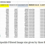
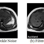
Speckle Noise is an inherent property of medical ultrasound imaging, and it normally tends to reduce the image resolution, pixel and contrast, thereby reducing the analytical value of the imaging modality. As a result, Speckle Noise reduction is an important prerequisite, whenever ultrasound imaging is used for tissue characterization [7]. As per the experiment, the best result on Speckle Noise was achieved by the Weiner filter [8]. First of the noise which was added was Speckle Noise to the 20 MRI images shown in the Fig 2(a). The noise which damages the quality of active radar, Medical Ultrasound, MRI images and Synthetic Aperture Radar is a granular noise which is known as Speckle Noise.
Filters used for the Speckle Noise were Weiner filter, median filter and Gaussian filter. The minimum size value for the filtered image is given by Weiner filter and clarity is also noted in fig 2 (b).
Blurred Noise
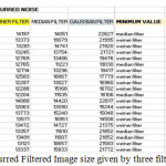
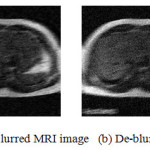
Second of the noise which is added to the images is Blurred Noise using function provided by MATLAB tool. J = imnoise (I,’n’,M,V) adds Blurred noise of noise n, mean m and variance v to the image I. The default is zero mean noise with 0.01 variance shown in the fig 4 (a). Blurred Noise is the noise which is present in the image that makes the image blurry, to remove this noise experimented filters are Gaussian filter, Median filter and Weiner filter. The minimum size values given by the filters after filtration are Weiner and Median filter but the clarity is noted by the Gaussian filter shown in the fig 4(b).
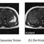
Gaussian Noise
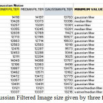
Third is the Gaussian Noise which is added to the images shown in the fig 6(a).Gaussian distribution which is also known as normal distribution whose probability is equal to statistical noise known as Gaussian Noise. Due to poor illumination and great extent of temperature or transmission of particles in an electronic image it fails to meet up the requirement for the clear image this noise is caused. By using a Weiner filter this noise can be reduced to very much extent. Same filters are used to check out the best filter for this noise. The best filter was Weiner filter but the minimum size value after using the different filters is given by all three filters shown in fig 5. Each filter is used on all the images, their outcome is noted and compared with the other entire filter applied on the same images. The circularly symmetric Gaussian behavior is found in the mellow ultra- sound speckle echo for marginal statistics which is similar to the laser speckle for monochromatic illumination [12].
The result which is achieved showed as follows:
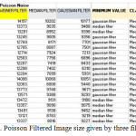
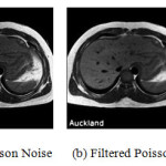
Poisson Noise
Poisson is also known as shot photon noise is the noise which is caused when sensor is not sufficient to provide detectable statistical information even after sensing number of photons [13]. This kind of noise is a type of electronic noise which occurs in an image due to small number of particles that carry energy [14]. This noise was added to the MRI images. Poisson distribution generally satisfies in many images which are having Poisson noise and also come across normally distributed and additive noise. Example radiography images and MRI images. Depending on the image intensity the magnitude of Poisson noise varies across an image which makes hard to remove the noise. All three filters Gaussian filter, Weiner filter, Median filter to remove Poisson Noise. The minimum size value after the filtration of the image was a contradiction between Gaussian and Median filter but the best clarity was achieved by Median filter with our experiment.
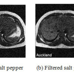
Salt Pepper Noise
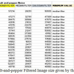
Fifth Noise is the Salt and Pepper Noise which is added to all the MRI images and the filtered applied on these images is Weiner Filter, Median Filter, and Gaussian Filter. Impulse noise is a kind of noise which is occurred due to the external channels and environment which affects the communication system, breakdown of image acquisition devices and sensor faults. Black and white dots which are usually seen in an image is due to this kind of digital images which are literally referred to as Salt-and-Pepper noise. The Median filter is used commonly to remove this kind of noise. In the experiment the used three filters were Weiner filter, Gaussian filter, Median filter [15] and the best result is observed by the median filter and the minimal size of an image was also noted by the same filter. A Super-Mean Filter (SUMF) has been used to remove high density Salt & Pepper noise from digital images. The working of this filter are generally in two stages, first stage is the detection of the noise in the image and the second stage is the pixel in an image is replaced by the mean value of the neighborhood noise free pixel[16].
Conclusion
In this work we have taken different medical images like MRI, and Brain for doing our experiment for noise removal. We have added Salt & Pepper, Gaussian, speckle, blurred and poison noise also removed these noises from the above medical images by applying the various filtering techniques like Median Filtering, Gaussian Filtering and Weiner Filtering techniques. In order to achieve accurate results for the given application it is mandatory to get good and clear images. The results are analyzed and compared with standard pattern of noises and also evaluated through the quality pixels, size, clarity and histogram of these images. Through this work we have observed that the choice of filters for de-noising the medical images depends on the type of noise and type of filtering technique, which are used. From the obtained result, on an average base of histogram, image size and clarity of the taken medical images Median filter gives best results for Poisson and Salt and Pepper noise. Weiner filter gives best results than all other filters for Gaussian and Speckle Noise. Gaussian filter give best results for Gaussian Noise images. Comparative results of all filters used for the noise are shown among all filtering methods based on image size, clarity and histogram. The results, which we have achieved, are more useful and they prove to be helpful for general to analyze which noised image can be de-noised using best filtering algorithm.
References
- Speckle Noise Reduction in Ultrasound Images- A Review Shruthi B, S Renukalatha, M SiddappaDept. of Computer Science and Engg. Sri Siddhartha Institute of Technology, Tumkur, Karnataka, India.
- Comparative Study of Fractional Filters for Alzheimer Disease Detection on MRI Images Samar M. Ismaila , Ahmed G. Radwanb,c, Ahmed H. Madianc,d, Mohamed F. Abu-ElYazeede a Faculty of IET, German University in Cairo (GUC), Egypt. b Dept. of Engineering Mathematics and Physics, Cairo University, Egypt. c NISC Research Center, Nile University, Cairo, Egypt. d Radiation Engineering Dept., NCRRT, Egyptian Atomic Energy Authority. e Electronics and comm. Eng. Dept., Cairo University, Egypt.
- Charles Boncelet (2005).”Image Noise Models”. in Alan C.Bovik. Handbook of Image and Video Processing.
- Research on Removing Noise in Medical Image Based on Median Filter Method NING Chun-yu 1.2, LIU Shu-fen’. QU Ming ‘ 1. Department ofComputer Science and Technology, Jilin University, Changchun, 130012, China; 2. School ofLife Science and Technology, Changchun University ofScience and Technology, Changchun, 130022, China.
- Enhancement Methods For Reduction Of Speckle In Ultrasound B- Mode Images 1 Kinita B Vandara, 2 DR. G. R. Kulkarni 1 Research Scholar, Department of Electronics and Communication, Shri J.J.T.University, vidyanagari, jhunjhunu, rajasthan 2 Principal, R.W.M.C.T’S Dnyanshree Institute of Engineering & Technology, Satara, Maharashtra.
- Noise Removal in Magnetic Resonance Images using Hybrid KSL Filtering Technique C.Lakshmi Devasena Department of Software Systems Karpagam Univeristy , M.Hemalata Department of Software Systems Karpagam Univeristy.
- Despeckling of Medical Ultrasound ImagesOleg V. Michailovich and Allen Tannenbaum, Member, IEEE.
- A Comparative Study on Approaches to Speckle Noise Reduction in Images Alenrex Maity, Anshuman Pattanaik, Santwana Sagnika, Santosh Pani School of Computer Engineering, Kalinga Institute of Industrial Technology, KIIT University Bhubaneswar,India.
- Comparative Analysis of Noise Removal Techniquesin MRI Brain Images B.Deepa Assistant Professor, dept of ECE Jayaram College of Engineering&TechnologyTrichy,India.Dr.M.G.SumithraProfessor, dept of ECE Bannari Amman Institute of Technology Erode, India.mgsumithra@rediffmail.com
- Performance Evaluation Of Filters In Noise Removal Of Finger Print Image 1 Ms.K.Kanagalakshmi, 2 Dr.E.Chandra1 Doctoral Research Scholar, 2 Director, Dept. of Computer Science, DJ Academy Managerial for Excellence.
- A Comparative Analysis of Filters on Brain MRI Images Neha Jain*, D S Karaulia Department of Computer Engineering and Application National Institute of Technical Teachers Training and Research Bhopal, India.
- Noise Through Removal of High Density Salt &Pepper Noise Through Removal of High Density Salt & Pepper Noise Through Super Mean Filter for Natural Images Super Mean Filter for Natural Images Super Mean Filter for Natural Images Super Mean Filter for Natural Images.
- Shyam Lal1, Sanjeev Kumar 2 and Mahesh Chandra3 1ECE Department, Moradabad Institute of Technology, Moradabad-244001(UP), India 2,3, ECE Department, Birla Institute of Technology,Mesra,Ranchi-835215(JH),India.
- A Comparative Study of Various Types of Image Noise and Efficient Noise Removal Techniques Mr. Rohit Verma Dr. Jahid Ali School of Information Technology SSCIMT, Badhani, Pathankot, APJIMTC, Jalandhar, India India
- Image De-noising by Various Filters for Different Noise Pawan Patidar Research Scholar (M. Tech.), Computer Science Department, Poornima College of Engineering, Jaipur, India, Sumit Srivastava Associate Professor, Computer Science Department, Poornima College of Engineering, Jaipur (Rajasthan), India.
- Study of Noise Detection and Noise Removal Techniques in Medical Images 1 Bhausaheb Shinde Head, Department of Computer Science, R.B.N.B. College, Shrirampur. Affiliated to Pune University Maharashtra, India Shinde.bhausaheb@gmail.com 2 Dnyandeo Mhaske Principal, R.B.N.B. College, Shrirampur. Affiliated to Pune University Maharashtra, India 3 A.R. Dani Head, International Institute of Information Technology, Hinjwadi, Pune Maharashtra, India.
- A Comparative Analysis of Filters on Brain MRI Images Neha Jain*, D S Karaulia Department of Computer Engineering and Application National Institute of Technical Teachers Training and Research Bhopal, India.

This work is licensed under a Creative Commons Attribution 4.0 International License.